| Revision as of 00:29, 19 September 2005 view sourceTony1 (talk | contribs)Autopatrolled, Extended confirmed users, Pending changes reviewers, Template editors276,546 edits →Prevention: Some minor edits and major queries.← Previous edit | Revision as of 00:37, 19 September 2005 view source Tony1 (talk | contribs)Autopatrolled, Extended confirmed users, Pending changes reviewers, Template editors276,546 edits →History of pneumoniaNext edit → | ||
| Line 94: | Line 94: | ||
| ==History of pneumonia== | ==History of pneumonia== | ||
| According to ''The Acorn'' newspaper (], ]), pneumonia was the leading cause of death in the ] in ].<!--Does it need a reference? It's kind of wordy and sounds parochial--> Before the discovery of ] in the late 1930s<!--check my guess-->, followed by other ], pneumonia was often fatal. <!--Meaning of 'first causal'? I've removed because now it doesn't fit; reinstate with different wording?-->Most community-acquired strains of ''S. pneumoniae'' are ] penicillin-sensitive. | |||
| Before the advent of ], pneumonia was often fatal. When ] was discovered in the ], it was the first causal therapy. Most community-acquired strains of ''S. pneumoniae'' are ] penicillin-sensitive. According to The Acorn Newspaper (], ]), pneumonia was the leading cause of death in the ] in ]. | |||
| ==See also== | ==See also== | ||
Revision as of 00:37, 19 September 2005
Pneumonia is an inflammation of the lungs. The term is almost always used to refer specifically to infections of the lungs caused by bacteria, viruses, fungi or other parasites; however, it can also refer to lung injury caused by physical or chemical irritants, in which case the term pneumonitis is used to differentiate the condition from infectious pneumonia. This article uses pneumonia only in the first sense, that of infection. Pneumonia may occur in people of all ages, although young children, the elderly, and immunocompromised patients are especially at risk. Antimicrobial drugs are often used to treat pneumonia.
Features
Symptoms of pneumonia commonly include shortness of breath; coughing that produces greenish or yellow sputum; a high fever (that may be accompanied with sweating, chills, and rigors ); sharp or stabbing chest pain, worsened by deep breaths or coughs (pleuritic chest pain); and rapid, shallow breathing that is often painful. Less commonly, there may be hemoptysis (the coughing up of blood), headaches (including migraine headaches), excessive sweating and clammy skin, loss of appetite, excessive fatigue, cyanosis (a blueness of the skin), nausea, vomiting, diarrhea, and arthalgia (joint pain) or myalgia (muscle aches). The manifestations of pneumonia, like those for many conditions, may not be typical in older people. They may instead experience new or worsening confusion, or falls.
Signs of pneumonia are tachypnea (rapid breathing), dullness to percussion, egophony, crackles, tachycardia (rapid heart rate), and fever.
Diagnosis
The differential diagnosis of pneumonia includes atelectasis, lung abscess, lung cancer, pulmonary embolism, and sepsis. To diagnose pneumonia, doctors rely on a patient's clinical history, findings from physical examination, and confirmatory scanning and pathology, typically including chest X-rays, blood tests, and sputum cultures. The chest X-ray is the usual standard for diagnosis in hospitals or clinics with access to X-ray facilities; however, in a community setting (general practice), doctors typically decide whether to start antibiotics for mild cases of pneumonia on the basis of a history and physical examination alone.
In nosocomial pneumonia (pneumonia that was acquired while the patient was in hospital for other treatment) and in immunocompromised patients, a clear diagnosis of pneumonia can be difficult; thus, a chest CT scan and/or other tests are often required to rule out causes such as pulmonary embolism). CT scanning may be useful when the symptoms and physical examination suggest several possible causes for the complaints (e.g., vasculitis, sarcoidosis, or lung cancer).
Physical examination
Apart from the history, the physical examination is an essential part of the doctor's overall assessment of the patient. Important features to note include whether the patient is breathless, able to speak in full sentences, uses accessory muscles of respiration, or has signs of reduced oxygenation (for example, blue, cyanotic lips, or unexplained mental confusion). If this overall assessment is poor, admission to hospital is usually advised, whatever else the examination reveals.
The pulse rate, repiratory rate and temperature are measured. Feeling for the expansion movements of the chest wall (palpation) and tapping the chest wall (percussion) to find resonant and dull areas may provide clues to the underlying disease process affecting the patient's lungs. Finally, auscultation with a stethoscope allows the doctor to listen for any areas of the lung which have reduced air flow, crackles (crepitus or 'rhonchi') or the crunch-sound (pleural rub) of pleurisy.

Chest X-rays, sputum cultures and other tests
The history and clinical examination often indicate the likelihood of the presence of pneumonia, and what alternative conditions may need to be ruled out. Depending on the setting, the severity of a patient's condition, the reliability of the diagnosis, and local conventions of medical practice, a doctor may consider arranging for laboratory testing and/or imaging to confirm the initial clinical data. The tests may include cultures of sputum and blood, a chest X-ray, and blood tests.
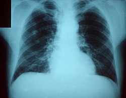
In the proper clinical setting, an increase in opacity in one or more lung fields on a chest x-ray, indicating consolidation of the infection in that region, helps to confirm the diagnosis of pneumonia. In community settings, radiologic studies can often take up to a fortnight to be interpreted. Chest X-rays are therefore only used by doctors practising in the community to investigate patients who are failing to respond to treatment, or who have easy access to chest radiography, such as an outpatient clinic run by a hospital. This is very different from the approach taken in the emergency room of a hospital, where the X-ray film is available for immediate viewing and therefore typically forms part of the initial investigations. Chest X-rays are not always accurate, either: they may miss pneumonias that can be seen on high-resolution computed tomography scans; they will miss pneumonias in which radiological signs have not yet developed, and they may result in the misdiagnosis of pneumonia when some other condition, such as pulmonary fibrosis or congestive heart failure, is responsible for the radiographic opacity. Opacities in the lower lobes are difficult to differentiate from atelectasis.
Should a doctor have any specific concerns about the diagnosis, or should a patient fail to recover after a course of antibiotics, a culture of the patient's sputum is normally requested. However, because it generally takes at least two days for a full analysis, sputum cultures are usually used only to retrospectively confirm the sensitivity of an infection to the antibiotic that has already been started. If possible, the culture should be collected prior to the start of antibiotics. The possibility of tuberculosis should be considered when a cough has been present for several weeks, or fails to respond to standard antibiotics. Special testing for tuberculosis needs to be specifically requested of the laboratory, because the bacterium that causes tuberculosis (Mycobacterium tuberculosis) cannot identified through the normal culturing process.
In the inpatient hospital setting, a blood sample is often routinely cultured to detect infection in the bloodstream (blood culture). As with sputum cultures, if the cultures grow bacteria, they can be identified and then tested to see which antibiotics will be most effective. Antimicrobial therapy can then be switched accordingly.
Supportive diagnostic tests usually include a full blood count; this may show a raised white cell count (neutrophilia), indicating the presence of an infection or inflammation (however, in some immunocompromised patients, the white cell count may appear deceptively normal). Renal function may have deteriorated if there is sepsis. There may be hyponatremia (low sodium levels), often due to the secretion of antidiuretic hormone by lung tissue; this is thought to be more frequent in tuberculosis and Legionaires' disease. Specific serological assays for atypical pathogens (Mycoplasma, Legionella and Chlamydia) are also available. In addition, there is now available a urine test for Legionella antigen.
Aetiology
There are over one hundred organisms known to cause pneumonia. However, relatively few are responsible for the majority of cases, and classifying the pneuomonias as mentioned above helps to shorten the list of likely offenders. Of these myriad pathogens, the most common cause of community-acquired pneumonia (the most common form of pneumonia overall) is Streptococcus pneumoniae. The major classes of microorganisms causing pneumonia are Gram-positive bacteria, Gram-negative bacteria, the so-called "atypical" bacteria, viruses, and opportunistic pathogens. This classification is important because different antimicrobial drugs are effective against different classes.
The major Gram-positive bacteria are Streptococcus pneumoniae, Staphylococcus aureus (often seen in more serious pneumonias, and Streptococcus pyogenes. Gram-negative bacteria are seen less frequently; more common pathogens include Haemophilus influenzae, Klebsiella pneumoniae, Pseudomonas aeruginosa, Moraxella catarrhalis, and Neisseria meningitidis. Escherichia coli, Proteus, and Enterobacter are responsible for a smaller portion of pneumonias. The atypical agents are Chlamydia pneumoniae, Mycoplasma pneumoniae, and Legionella pneumophila.
Viral pneumonia is usually caused by influenza virus, respiratory syncytial virus (RSV), adenoviruses, and varicella-zoster virus (direct VZV pneumonia is rare, but well recognosied is a staphylococcal secondary infection in cases of chicken pox. Herpes simplex virus is a rare cause of pneumonia. The above agents all cause pneumonia in people with intact immune systems. In those whose immune system is impaired (for instance, due to infection with HIV or as a result of taking immunosuppresive drugs), opportunistic pathogens can cause pneumonia. Major ones include the Pneumocystis jiroveci (a fungus), Mycobacterium avium (a bacterium), and cytomegalovirus (CMV, a virus).
Types of pneumonia
There are several different classification schemes: microbiological, radiological, age-related, anatomical, point of acquiring infection. The main classification used in medical journals is that between the point of infection: community-acquired and hospital-acquired. Community-acquired pneumonias are pneumonias in a patient who is not or has not recently been hospitalized, whereas hospital-acquired pneumonias (or nosocomial pneumonias) are found in hospitalized or recently discharged patients. Furthermore, infections in the immunocompromised, as well as aspiration pneumonia, are usually treated as separate disease entities as they have other causal agents, as well as a different clinical course.
Aside from these, there are several other terms used to classify pneumonias. A lobar pneumonia is an infection that involves, and is limited to, a single lobe of a lung (generally due to Streptococcus pneumoniae). In contrast, multilobar pneumonia involves more than one lobe. Ventilator-associated pneumonia can be considered a subset of hospital-acquired pneumonia; it occurs following intubation and mechanical ventilation for at least 48 hours. "Walking pneumonia" is an outdated term for pneumonia in a patient who is still able to walk—that is, a mild pneumonia, usually due to Mycoplasma. Pneumococcal pneumonia is due to S. pneumoniae (around half of all pneumonias). Finally, atypical pneumonia is due to either Mycoplasma, Chlamydia, or Legionella.
Community-acquired pneumonia
Epidemiology
Community-acquired pneumonia (CAP) is a serious illness. It is the fourth most common cause of death in the UK, and sixth in the USA. 85% of cases of CAP are caused by the typical bacterial pathogens, namely, Streptococcus pneumoniae, Haemophilus influenzae, and Moraxella catarrhalis. The remaining 15% are caused by atypical pathogens, namely Mycoplasma pneumoniae, Chlamydia pneumoniae, and Legionella species. Unusual aerobic gram-negative bacilli (for example, Pseudomonas aeruginosa, Acinetobacter, Enterobacter) rarely cause CAP.
Clinical features
Typical symptoms include cough, purulent sputum production, shortness of breath, pleuritic chest pain, fevers and chills. On examination, one notes rapid respiratory rate and heart rate and signs of pulmonary consolidation. In the elderly, symptoms and signs are sometimes vague and non-specific. They may include headache, malaise, diarrhea, confusion, falling, and decreased appetite. Diagnosis is confirmed by physical examination and chest x-ray. In general, patients who present with symptoms consistent with CAP, without extrapulmonary findings on history, physical examination or in laboratory tests have a CAP caused by a typical pathogen. Patients who have pneumonia plus extrapulmonary physical findings or laboratory features (such as elevations in liver function test results) have an atypical pneumonia.
Hospital-acquired pneumonia
Hospital-acquired pneumonia, also called nosocomial pneumonia, is a lung infection acquired after hospitalization for another illness or procedure. It is considered a separate clinical entity from CAP because the causes, microbiology, treatment and prognosis are different. Up to 5% of patients admitted to an hospital for other causes subsequently develop a pneumonia. Hospitalized patients have a variety of risk factors for pneumonia, including mechanical ventilation, prolonged malnutrition, underlying cardiac and pulmonary diseases, achlorhydria and immune disorders. Additionally, pathogens thrive in hospitals that could not survive in other environments. These pathogens include resistant aerobic gram-negative rods, such as Pseudomonas, Enterobacter and Serratia, resistant gram positive cocci, such as MRSA. Because of risk factors, underlying morbidity and resistant bacteria, hospital-acquired pneumonia tends to be more deadly than its community counterpart.
Ideal therapy is based on determination of the aetiological agent and its relevant antibiotic sensitivity; however, a specific pathogen is identified in only 50% of patients even with extensive evaluation. Empiric treatment is usually started before laboratory microbiological reports are available as treatment should not be delayed in any patient due to the seriousness of the disease.
Antibiotics used for hospital-acquired pneumonia include aminoglycosides, fluoroquinolones, carbapenems, and vancomycin. Multiple antibiotics are administered in combination in order to cover all the possible organisms effectively and rapidly, before the infectious agent can be known. Antibiotic choice varies from hospital to hospital as the likely pathogens and resistance patterns vary from place to place.
Other pneumonias
- Severe acute respiratory syndrome (SARS)
- Pneumocystis carinii pneumonia
- Bronchiolitis obliterans organizing pneumonia
- Eosinophilic pneumonia
- Chemical pneumonia, or more properly chemical pneumonitis, caused by chemical toxins such as pesticides or gasoline which enter the body by inhalation or by skin contact.
Pathophysiology
The vast majority of pneumonias are infectious diseases; whether a patient is prone to develop pneumonia depends not only the presence of pathogens but equally on the patient's immune system and other factors. Most pneumonias are not epidemic, although infection with influenza virus can be so defined.
Breathing problems, as often present in patients after a stroke, in Parkinson's disease, hospitalisation or surgery and mechanical ventilation can all increase the likelihood of pneumonia. Similarly, inability to clear sputum (as in cystic fibrosis) or retention of sputum (as in bronchiectasis) can lead to pneumonia.
After splenectomy (removal of the spleen), a patient is more prone to pneumonia due to the spleen's role in developing immunity against the polysaccharides on pneumococcus bacteria.
Therapy
Antibiotics are the treatment of choice for bacterial pneumonia. They are not effective in viral pulmonary infections, but are sometimes used against concommitant bacterial superinfection. The choice of antibiotic depends on the nature of the pneumonia, the microbes known typically to cause pneumonia in the geographical region, and the immune status of the patient. In the United Kingdom, Amoxicillin is used as first-line therapy in the vast majority of patients who acquire pneumonia in the community, sometimes with added clarithromycin. In North America, where the "atypical" forms of community-acquired pneumonia are becoming more common, clarithromycin, azithromycin, and the fluoroquinolones have displaced the penicillin-related drugs as first-line therapy. In hospitalized patients and immune-deficient patients, local guidelines determine the selection of generally intravenous) antibiotics.
Patients who have significantly compromised respiratory function due to pneumonia may require supplemental oxygen. Severely affected patients may require artificial ventilation as a life-saving measure while their immune system fights off the infective cause with the help of antibiotics and other drugs. In cases of viral (interstitial) pneumonia where influenza A or B are thought to be causative agents, patients who are seen within 48 hours of the onset of symptoms may be treated with oseltamivir or zanamivir. There is no known effective treatment for pneumonia caused by SARS coronavirus, adenovirus, hantavirus, or parainfluenza virus.
Complications
- sepsis
- lung abscess or empyema
- pleural effusion, pleuritis
- acute respiratory distress syndrome (ARDS), respiratory failure
- pneumothorax (rare)
Prognosis and mortality
The clinical state of the patient at time of presentation is a strong predictor of the clinical course. Many clinicians use the Pneumonia Severity Score to calculate whether a patient requires admission to hospital, based on the severity of symptoms, underlying disease and age![]() Administrator note. In the United States, mortality from pneumococcal pneumonia is 1 in 20. In cases where the disease progresses to blood poisoning (bacteremia), 2 of 10 die. When the disease affects the brain (meningitis), 3 of 10 die.
Administrator note. In the United States, mortality from pneumococcal pneumonia is 1 in 20. In cases where the disease progresses to blood poisoning (bacteremia), 2 of 10 die. When the disease affects the brain (meningitis), 3 of 10 die.
Prevention
Vaccination with the pneumococcal polysaccharide vaccine is recommended for adults older than 65, patients with chronic disease, and patients with immune compromise (including HIV, nephrotic syndrome and asplenia). Pneumoccocal pneumonia kills more Americans than all other diseases combined that could be partially prevented by vaccination.
These groups should also have annual flu vaccination to avoid secondary bacterial infections after influenza infection.
Epidemiology
Pneumonia is mainly a disease of young children and the elderly. Young adults rarely get the disease. It occurs more often during winter and spring than during summer and autumn.
History of pneumonia
According to The Acorn newspaper (Conejo Valley, Southern California), pneumonia was the leading cause of death in the United States in 1904. Before the discovery of penicillin in the late 1930s, followed by other antibiotics, pneumonia was often fatal. Most community-acquired strains of S. pneumoniae are still penicillin-sensitive.
See also
References
- Template:Anb Halm EA, Teirstein AS. Management of community-acquired pneumonia. N Engl J Med. 2002;347:2039-45. PMID 12490686.